
Cell Cycle Chromobody Immunodetection Vector
Antibody Detection within Living Cells
A non-invasive way to trace cell cycle dynamics in live cells
A new tool for detection of PCNA in live cells, the Cell Cycle Chromobody, lets you monitor the cell cycle as it happens. The small size of the Chromobody produces a high signal to noise for stunning, high resolution images or time lapsed movies clearly show the punctate pattern of replication sites within the nucleus of actively dividing cells. Simply transfect the cell line of your choice with the Cell Cycle Chromobody plasmid. This plasmid contains the sequence of an PCNA-specific alpaca antibody fused to TagRFP (Evrogen). PCNA is known to be a scaffolding protein which helps anchor and recruit DNA processing enzymes to the appropriate sites throughout the chromatin. Upon transformation, your cells will express the Cell Cycle Chromobody. The transient binding does not influence cell viability or progression through the phases of the cell cycle, and thus offers you a unique possibility to observe your cell’s growth state.
U2OS cell line stably expressing Cell Cycle Chromobody fused to the red fluorescent protein TagRFP
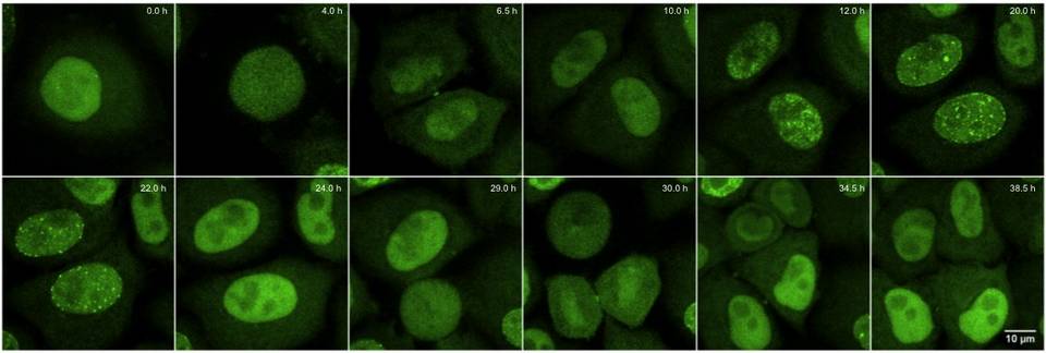
The above panel shows how the Cell Cycle Chromobody-TagGFP2 [No longer available] clearly follows a cell over a 36 hour time course in which it undergoes 2 rounds of division. After the first division, the daughter cells enter S-Phase around 10 hours showing characteristic punctate pattern of PCNA. The 29 hour mark shows one of the daughter cells entering Mitosis (prometphase), with the second following closely behind at hour 30. For live cell imaging, cells were seeded in ibidi µ-slides. Time series was acquired on a spinning disk microscope (UltraVIEW VoX, PerkinElmer) equipped with a 63x/1.4 NA Plan-Apochromat oil immersion objective and a humidified, temperature- and CO2-controlled live cell chamber. TagGFP2 was excited with a 488 nm laser-line for typically between 100-500 ms/ image with laser intensity set to max. 25%. Confocal image z-stacks of living cells were recorded with a frame size of 1000×1000 pixels, a pixel size of 100 nm and a z-step size of 400 nm every 30 minutes. Images shown are maximum intensity projections of few mid-z-sections.
Chromobodies turn your cells into immuno-reporter factories
Nanobodies are unique, small antibody fragments that exhibit the extraordinarily efficacious binding of a targeted protein. Expressed in animal cells, Nanobodies can be collected and used for in vitro studies. Something unexpected happened when Nanobodies were expressed as a fusion to a reporter molecule such as GFP. The newly formed Chromobody bound directly to its target within the cells in which it was being made. Moreover, using the correct vector and promoters, the binding has both high specificity and low background. This provides an excellent alternative to GFP-fusions with the protein of interest for immuno-localization studies. Why? Because GFP- and RFP-fusions have been shown to block or alter the activity of the fused protein. In contrast, Chromobody act in trans as a reporter that will not interfere with the normal function of the targeted protein.
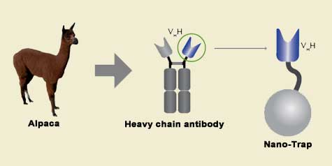
Camelidae single-domain antibodies are like IgGs on steroids
The family of animals known as Camelidae (camels, llamas, and alpacas) produce functional antibodies devoid of light chains, so-called "heavy chain" antibodies. These heavy chain antibodies recognize and bind their antigens via a single variable domain. When cleaved from their carboxy tail, these barrel-shaped structures (2x3 nm) are extraordinarily small, naturally-occurring, and intact antigen binding fragments (MW of 13 kDa). These fragments, called Nanobodies, are characterized by high specificity, affinities in the low nanomolar range, and dissociation constants in the sub-nanomolar range (typically 10- to 100-fold better than mouse IgGs). The compact size of Nanobodies makes them extremely stable at temperatures up to 70°C, and functional even in 2M NaCl or 0.5% SDS. These small and powerful antibody fragments can be used in a variety of unique applications. They will open up your research possibilities.
Cell Cycle Chromobody Immunodetection Vector
|
SPECIFICATIONS
|
|
|---|---|
| Part Numbers | CCBRFP, ccg, ccr |
| Vector Type | Mammalian expression vector encoding the cell cycle marker PCNA-VHH fused to green fluorescent protein TagRFP |
| Target Molecule | PCNA |
| Reporter | TagRFP (from Evrogen) |
| Host Cells | Mammalian |
| Transfection Method | Transfect mammalian cells by any known transfection method. If required, stable transformants can be selected using G418 [Gorman 1985] |
| Propagation in E. coli | DH5alpha, HB101, XL1-Blue, and other general purpose strains
Incompatible with pMB1/ColE1 |
| Selection | Prokaryotic – kanamycin
Eukaryotic – neomycin (G418) |
| Replication | Prokaryotic – pUC ori
Eukaryotic – SV40 ori |
| Use | Non-invasive live cell visualization of cell cycle progression |