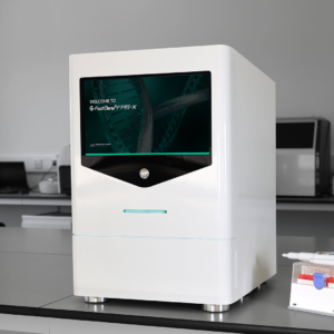
FAS-X Gel Imaging System
Advanced Gel Documentation with Unique Blue/Green LED Light
Perfect results right out of the box
The FastGene FAS-X Gel Imaging system is the latest addition to the growing FastGene family for sensitive and safe nucleic acid detection and protein documentation. This imaging platform starts with a powerful CMOS 20 megapixel camera and combines that with the superior technology of ultra-bright blue/green LEDs for luminous imaging of nearly any fluorescent dye. These brilliant images are easy to access on a large touch-screen display. The aquamarine-color illumination emitted by these special blue/green LEDs is different from the monochromatic color of your typical blue LEDS as it hits the “sweet spot” for maximum excitation of common fluorescent DNA dyes such as EtBr and Sybr Green. Expect equivalent or better results as compared to UV transilluminators, but without the risk of damaging DNA or harming your skin and eyes. That’s because the lower energy photons from blue/green LEDs will not crosslink DNA (rendering it unable to be amplified by PCR) unlike short wave UV light.
The FAS-X system is designed to produce stronger signals and lower background! The huge imaging area is 26 cm x 21 cm and has unsurpassed uniformity of lighting. 12 LED arrays illuminate from opposite sides (trans-lighting) as compared to directly shining light from below like other gel illuminators. The FAS-X was developed for use with either red dyes such as ethidium bromide (EtBr) or next generation non-carcinogenic dyes like our Midori Green or Sybr Green-based dyes.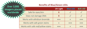

Blue/Green Illumination vs UV Illumination
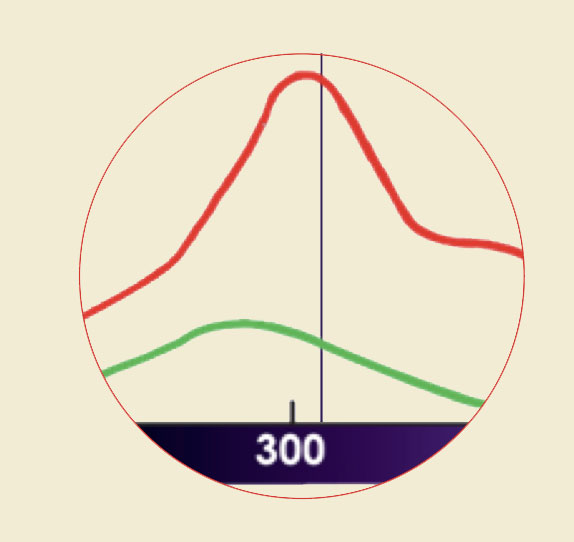 UV-Light: good signal, unhealthy side-effects
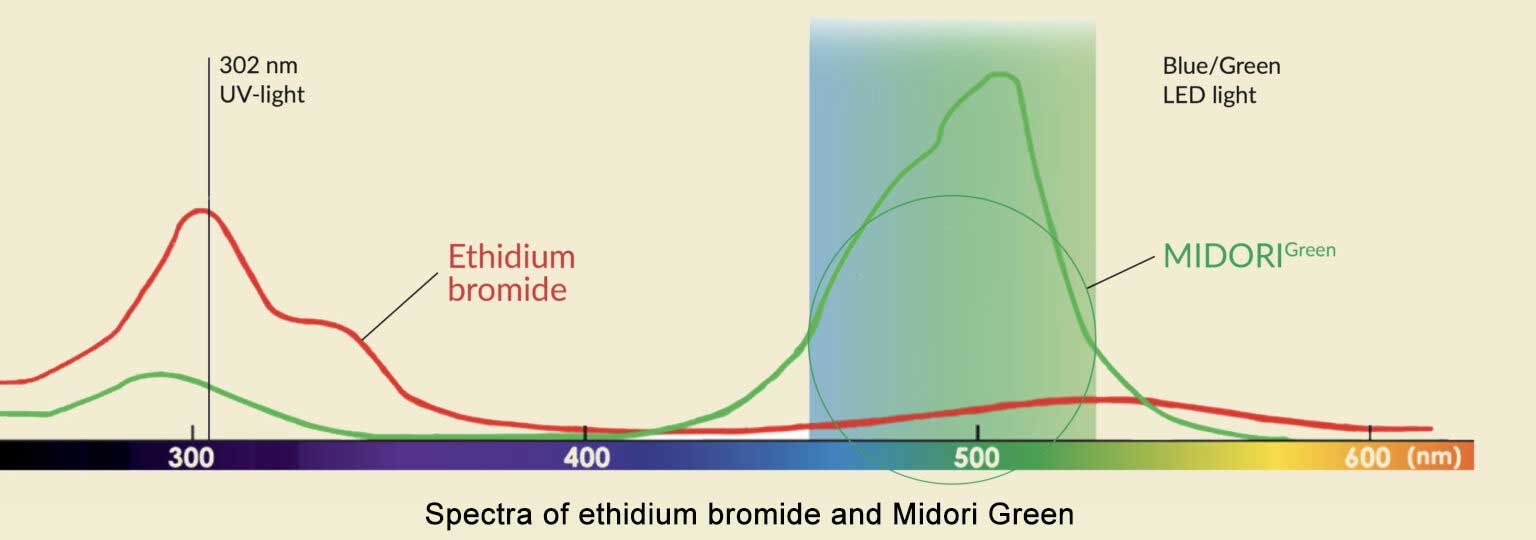
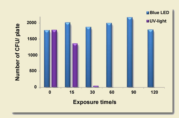
UV-light transilluminators use a single wavelength for the visualization of DNA. Red and green DNA dyes, such as ethidium bromide or Midori Green dyes have a good absorption in the UV-light spectrum. This results in DNA bands with sufficient intensity, however, UV-light is dangerous for the user and for the sample DNA. Just 30 seconds of UV-light exposure significantly reduces the cloning efficiency and has consequences for downstream applications. For this reason, visualization of DNA with UV-light is not the preferred method.

Images of different gel stains with Blue/Green illumination
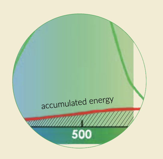 Blue/Green (not Blue) LEDs: safe and strong detection of all DNA dyes
In contrast to UV-light, Blue/Green LED technology uses a wide spectrum of light between 470 nm and 520 nm. This range of wavelengths is much broader than emitted by blue LEDs. This light is not harmful for DNA or for the user. Even ethidium bromide or other red DNA dyes with a low absorption in this spectral area show DNA band intensity comparable to UV illumination. The reason for that is the accumulated energy absorption (area under the curve) of the DNA in the Blue/Green spectrum. Green DNA dyes show very high absorption intensity in the Blue/Green light spectrum, leading to DNA bands with superior intensity.
Touch-screen operation
The FastGene FAS-X is easily controlled by a gorgeous, color 13.3″, high resolution, touchscreen display. Direct communication between the software and the camera enables fast image capture and powerful live image editing.
Auto or Pro Mode – Only 3 taps to get the perfect gel image
The AUTO mode automatically optimizes the image capture process for you. It allows users to obtain the finest gel images with just a few taps.
- Turn on Blue/Green LED lamps
- Adjust the brightness
- Capture the image

The PRO mode gives you full control over the image capture settings. You can fine-tune the brightness, zoom in and change the color scheme (color, monochrome and inverted colors) to get the image you want. The functionality of PRO mode even extends to multi-image recording with 3 or 5 images recorded with different exposure times. This allows you to quickly select the perfect shot.

Many illumination options for different experiments
The FAS-X system comes with blue-green and white light illumination on-board:
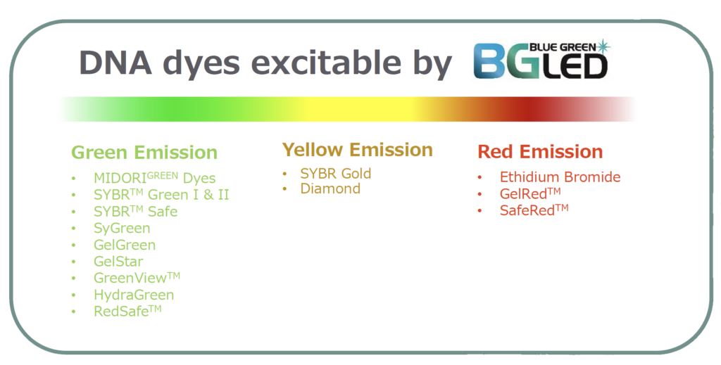
Blue-Green Illumination – Blue/Green LED technology enables the detection of red and green DNA dyes such as Midori Green, Sybr Green and Ethidium Bromide (EtBr). This mode is most commonly used for the detection of DNA and RNA (both single and double stranded).
|
Green fluorescent dyes
|
Red fluorescent dyes
|
 |
 |
MIDORI Green Xtra DNA dye |
GelRed DNA dye |
 |
 |
SYBR Safe DNA dye |
SERVA PRiME Lightning Red |
White Illumination – High-resolution images of (colorimetric) western blot membranes or bacterial colony plates can be obtained in white light mode. Together with the included white plate, the white light mode can be used to visualize colourimetric PAGE protein gels with an impressive quality.


Safer for you and better for your subcloning
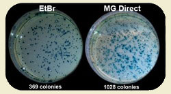
By using safe MIDORI Green dyes and safe Blue/Green LED illumination you can improve your subcloning transformation efficiencies by THREE-FOLD. In the example below, a plasmid vector was double digested with suitable restriction enzymes to create two sticky-ended DNA fragments: the lacZ gene (3,536 bp) and the backbone of the vector (4,318 bp). Equal amounts of digested DNA were electrophoresed on 1% agarose gels. The gels were stained with either ethidium bromide or MIDORI Green Direct gel stain according to the corresponding manuals, and then viewed using either a UV transilluminator or the FastGene Blue/Green LED Illuminator, respectively. The two DNA fragments were excised from the gels and purified using a silica membrane based purification kit. The lacZ gene and the vector backbone were re-ligated using T4 DNA ligase transformed into DH5a cells and plated onto selection plates. The total number of blue and white colonies was counted to evaluate cloning efficiency. Each experiment was conducted in triplicate, and the average cloning efficiency was determined. MIDORI Green Direct resulted in a dramatic increase of positive transformants.
Midori Green Can Boost Your Cloning Results!
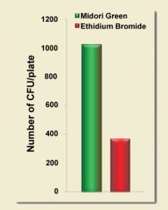
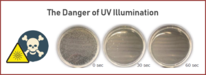
Ethidium bromide is typically used in conjunction with a strong UV light source to excise DNA bands for purification prior to the ligation reaction. Short-wavelength light is known to cause thymine dimers and damage DNA. The extent of this damage is not always appreciated. High-energy light wreaks havoc on DNA fragments in mere seconds. As can be seen below, cloning efficiency starts to drop after just a 15 second exposure of DNA in a standard agarose gel. After a 30 second exposure, your cloning experiment is all but dead! In contrast, the cloning efficiency of protocols that use blue LEDs or Nippon Genetics’ super-performing Blue/Green LEDs are completely unaffected by this deleterious effect. If your lab can’t to break itself of its ethidium bromide habit, using a Blue/Green LED Illuminator (or imaging system) should still have an immediate positive impact on DNA integrity and cloning efficiency.
UV Transilluminators Kill Cloning Experiments
FAS-X Gel Imaging System
FAS-X Accessories
|
SPECIFICATIONS
|
|
|---|---|
| Applications | DNA/RNA
-All Green and Red fluorescent dyes such as Sybr Green, Midori Green & GelGreen, EtBr & GelRed, ext. PROTEIN GELS & BLOTS – SYPRO Ruby, Coomassie PETRI DISH – Documentation and fluorescence |
| Part Numbers | GP-FAS-X |
| Fluorescence | Yes |
| Colormetric | Yes |
| Gel Documentation | Yes |
| Excise Bands | Yes |
| Light Sources | Blue/Green LED transilluminator (built-in light), White LED room light (integrated Epi white light) White LED transilluminator (external white light plate supplied) |
| Blue/Green Light Specs | 470 – 520 nm |
| Filter | Amber camera filter, amber goggles included |
| Camera Sensor | Scientific-grade 1″ CMOS |
| Image Resolution | 20 MPixel (5472 px x 3648 px) |
| Image Format | JPEG, TIFF, PNG, BMP |
| Exposure time | 13 µs to 10 sec |
| Imaging Area | 26cm x 21cm |
| Touchscreen | 13” full HD glove compatible touchscreen |
| Display Resolution | 1920 px x 1080 px |
| Connections | LAN, 3 x USB 3.0 |
| Network Compatible | Yes |
| Storage | Internal 128GB |
| Size | 37.5 cm x 44.3 cm x 53.2 cm x (Length x Width x Height) |
| Image Type | TIFF, JPEG, BMP & PNG |
| Image Editing | Onboard image editing |
| Security | User login and password protection |
| Weight | 20 kg |
| Power | 24V, 6A, 100-240V, 50-60Hz |
| Warranty | 1 year |


