FastGene FAS-BG LED BOX
Gel Documentation System for Every Day & Every Dye
High-end gel documentation at a low-end price
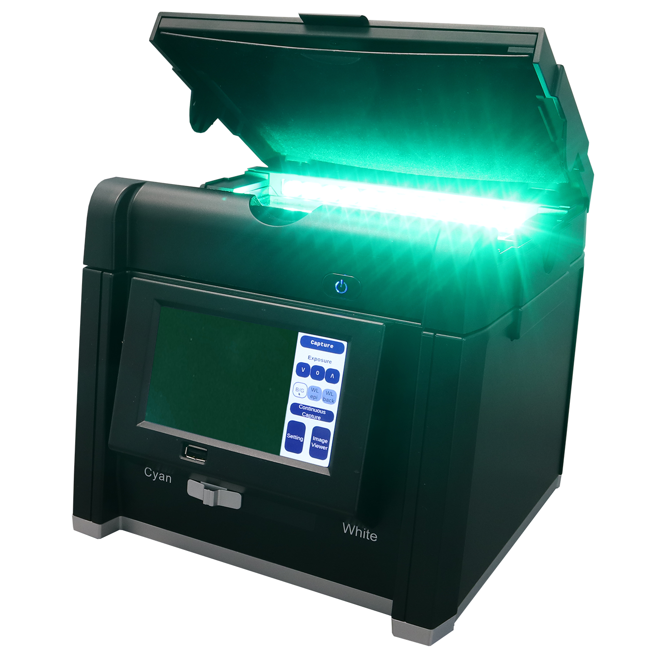
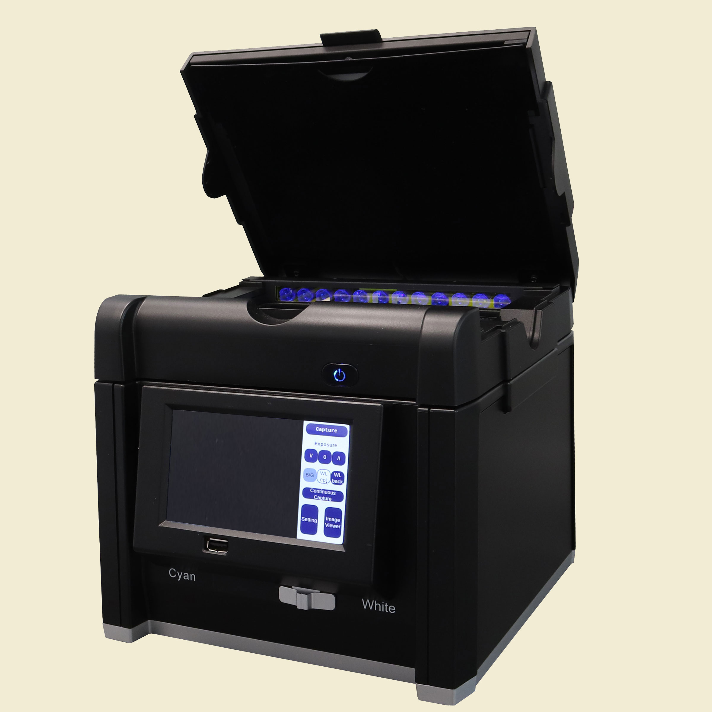
The FastGene FAS-BG LED BOX is the most compact gel documentation systems available. Ideally suited for tight spaces on any bench-top, the system both illuminates and captures an image of your DNA, RNA, or protein gel. The FAS-BG LED BOX boasts a large touchscreen interface and comes equipped with three different illumination modes for even more uses.
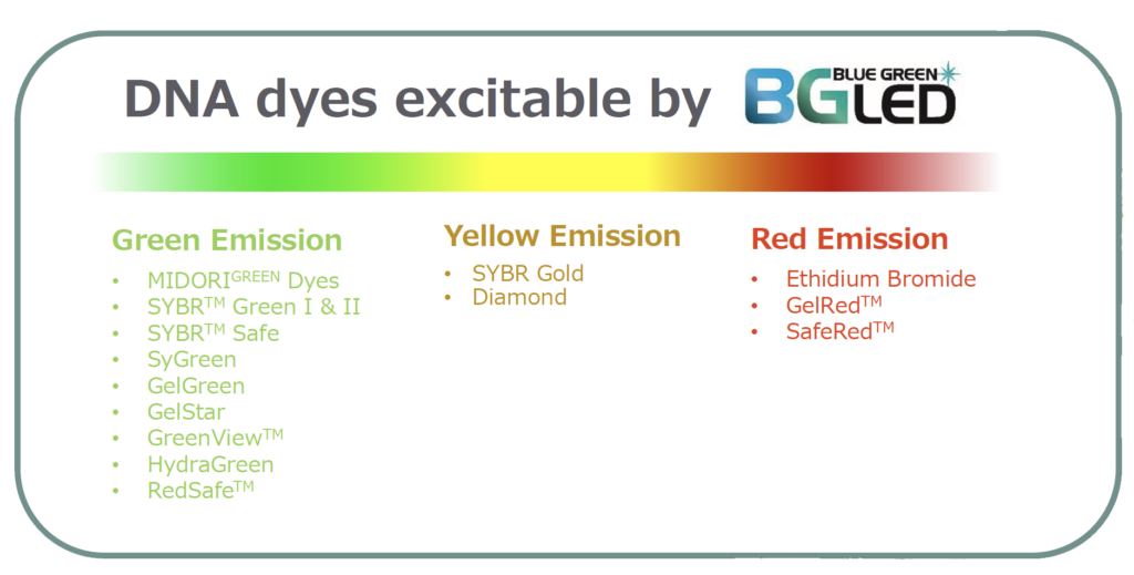
Blue/Green Mode: An array of blue/green LEDs (515 nm) situated around the periphery of the glass platen provide excitation light for both red- and green–emitting fluorescence dyes. Now you can get great images with traditional EtBr, an impossible feat with “blue-only” LED illuminators! This optimal “transillumination” configuration minimizes ambient background for super sensitive imaging with non-carcinogenic dyes like Midori Green, too. Most importantly, blue/green LEDs are just as sensitive as UV light but don’t damage DNA in an agarose gel—and they won’t harm your skin or your eyes.
Trans White Mode: An innovatively-designed white LED array shines uniform backlight for transparent media such as Coomassie-stained protein polyacrylamide gels. This diffuser technology eliminates “hot spots” due to point illumination and provides maximum clarity for colorimetric-stained gels.
Epi White Mode: The final mode uses white LED “epi-illumination” for lighting from above. This mode is suited for opaque surfaces such as Western blots and similar highly reflective materials.
No matter the illumination mode, the FAS-BG LED BOX system captures images with a high-end, scientific grade, 8 megapixel CMOS camera for wonderful separation of color and resolution of bands. These images can be saved to a computer, or you can attach a printer directly for quick printouts.
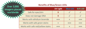
Blue/Green Illumination vs UV Illumination
 UV-Light: good signal, unhealthy side-effects
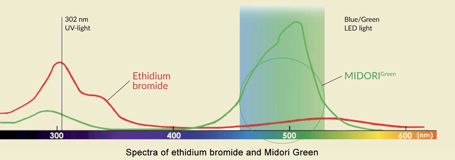
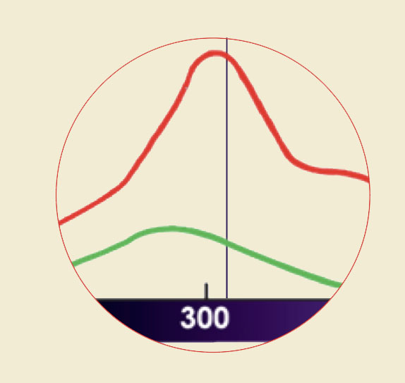
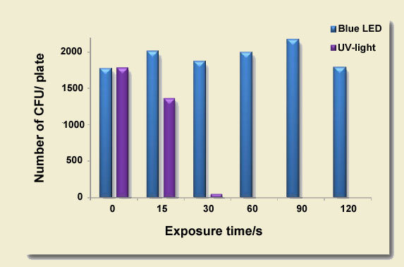
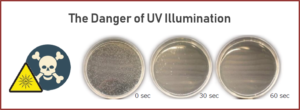
UV-light transilluminators use a single wavelength for the visualization of DNA. Red and green DNA dyes, such as ethidium bromide or Midori Green dyes have a good absorption in the UV-light spectrum. This results in DNA bands with sufficient intensity, however, UV-light is dangerous for the user and for the sample DNA. Just 30 seconds of UV-light exposure significantly reduces the cloning efficiency and has consequences for downstream applications. For this reason, visualization of DNA with UV-light is not the preferred method.

Images of different gel stains with Blue/Green illumination
 Blue/Green (not Blue) LEDs: safe and strong detection of all DNA dyes
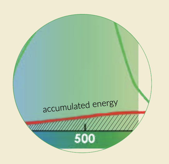
In contrast to UV-light, Blue/Green LED technology uses a wide spectrum of light between 470 nm and 520 nm. This range of wavelengths is much broader than emitted by blue LEDs. This light is not harmful for DNA or for the user. Even ethidium bromide or other red DNA dyes with a low absorption in this spectral area show DNA band intensity comparable to UV illumination. The reason for that is the accumulated energy absorption (area under the curve) of the DNA in the Blue/Green spectrum. Green DNA dyes show very high absorption intensity in the Blue/Green light spectrum, leading to DNA bands with superior intensity.
Integrated Touchscreen
FastGene FAS-BG LED BOX comes with a 5″ touchscreen display and built-in software which allows for easy navigation and image capture. For those looking for a larger display, there is an HDMI port which can easily be connected to an external display or touch monitor. And for those looking for a familiar feel, the FAS-BG LED BOX display can be controlled by a USB mouse with or without an external monitor.

Excise your DNA fragments the easy way
The FastGene FAS-BG LED BOX comes with an amber filter shield that can be fastened on the upper lid.
The amber filter shield filters out the Blue/Green LED excitation wavelengths and allows a clear visualization of DNA bands.

Documentation of petri dishes, protein gels and Western Blot images
- Colorimetric protein gels can be visualized with white LED light.
- The white epi light is used fo the document opaque surfaces such as Petri dishes or membranes with

Safer for you and better for your subcloning
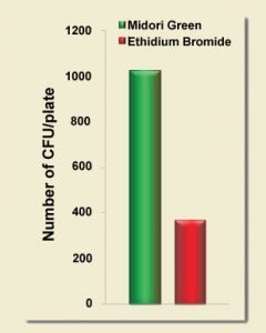
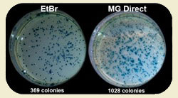
By using safe MIDORI Green dyes and safe Blue/Green LED illumination you can improve your subcloning transformation efficiencies by THREE-FOLD. In the example below, a plasmid vector was double digested with suitable restriction enzymes to create two sticky-ended DNA fragments: the lacZ gene (3,536 bp) and the backbone of the vector (4,318 bp). Equal amounts of digested DNA were electrophoresed on 1% agarose gels. The gels were stained with either ethidium bromide or MIDORI Green Direct gel stain according to the corresponding manuals, and then viewed using either a UV transilluminator or the FastGene Blue/Green LED Illuminator, respectively. The two DNA fragments were excised from the gels and purified using a silica membrane based purification kit. The lacZ gene and the vector backbone were re-ligated using T4 DNA ligase transformed into DH5a cells and plated onto selection plates. The total number of blue and white colonies was counted to evaluate cloning efficiency. Each experiment was conducted in triplicate, and the average cloning efficiency was determined. MIDORI Green Direct resulted in a dramatic increase of positive transformants.
Midori Green Can Boost Your Cloning Results!
Ethidium bromide is typically used in conjunction with a strong UV light source to excise DNA bands for purification prior to the ligation reaction. Short-wavelength light is known to cause thymine dimers and damage DNA. The extent of this damage is not always appreciated. High-energy light wreaks havoc on DNA fragments in mere seconds. As can be seen below, cloning efficiency starts to drop after just a 15 second exposure of DNA in a standard agarose gel. After a 30 second exposure, your cloning experiment is all but dead! In contrast, the cloning efficiency of protocols that use blue LEDs or Nippon Genetics’ super-performing Blue/Green LEDs are completely unaffected by this deleterious effect. If your lab can’t to break itself of its ethidium bromide habit, using a Blue/Green LED Illuminator (or imaging system) should still have an immediate positive impact on DNA integrity and cloning efficiency.
UV Transilluminators Kill Cloning Experiments
FastGene FAS-BG LED BOX
Accessories
|
SPECIFICATIONS
|
|
|---|---|
| Applications | DNA/RNA
-Green fluorescent dyes such as Sybr Green, Midori Green & GelGreen, ext. -Red fluorescent dyes such as EtBr & GelRed, ext. -Petri dish documentation -Western Blot images (colormetric only) |
| Part Numbers | GP-04LED |
| Wavelength | 470-520nm Blue/Green LEDs |
| Filter | Included amber filter shield |
| LED Array | Dual matrices for two-sided illumination |
| LED Lifetime | 50,000 hours |
| Camera | 8 Mega Pixel camera with CMOS sensor |
| Exposure time | 0.2 – 2 sec, 21 different exposure settings |
| Display | 5” color LCD touch panel |
| Software | On-board control software |
| Connections | USB 2.0 port (1x front, 1x back)
1x HDMI port Thermal printer support |
| Size | 23 x 26 x 21 cm (length x height x depth) |
| Illumination Surface | 16 x 11.5 cm |
| Image Type | TIFF, JPEG and PNG |
| Weight | 3.2 kg |
| Power | 100-240V, 50-60Hz |
| Warranty | 1 year |

