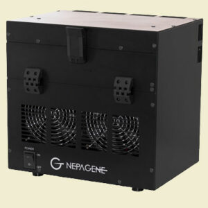
TiYO Autofluorescence Quenching System
Eliminates sample autofluorescence for microscopy
Maximize signal and minimize background with no chemical treatment
The TiYO Autofluorescence Quenching System is the first-of-its-kind instrument to help eliminate the background fluorescence signal that is common in most cell and tissue types. Autofluorescence is commonly derived from lipofuscin, elastin fibers, vitamin A, and other intrinsic molecules, and can often make fluorescence imaging of tissues difficult. The reduction of fluorescence background leads to more precise signal localization when using immunofluorescence or fluorescent cell staining techniques. The TiYO works by both photo-bleaching the unwanted autofluorescence and also maintaining samples at an ambient temperature during this process so that no tissue degradation occurs.

Simple to use design
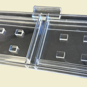
The TiYO coudn’t be any easier to use. Simply place between 1 and 12 samples in the tray, insert in TiYO, and then expose for 30 minutes to 2 hours depending upon the tissue type. Once complete, the samples are now ready for normal processing for whichever fluorescence staining technique your experiment requires. Unlike conventional quenching reagents, with the TiYO only the autofluorescence signal is affected. In other words there is no reduction in the fluorescence intensity from stains that are applied after TiYO treatment.

Comparative data of fluorescence staining without treatment, with TiYO™ pretreatment, and with reagent treatment.
From the images below you can see that the TiYO™ (the middle panels) has little effect on the fluorescence signal derived from stains. While the traditional quenching reagent directly reduces the intensity of fluorescent stains:

Effective for high autofluorescence tissue types
The image demonstrates the quenching effect using TiYO with chum salmon gill sections*. Chum salmon gill sections are known for their extraordinarily high levels of autofluorescence. A simple two hour treatment with the TiYO eliminated the autofluorescence (the same imaging conditions and contrast settings were used for all four images)
*Courtesy of Dr. Takehiro TSUKADA / Department of Biomolecular Science, Faculty of Science, Toho University

Reveal hidden signals
Sometimes important staining data is hopelessly lost in the noise. The TiYO can uncover these structures and patterns, such as demonstrated here. In this case, mouse brain tissue sections have unusual ring-shaped structures similar in size and shape as the staining target. It is not until after the tissue sections are quenched with the TiYO system that the interesting results are revealed. (the same imaging conditions and contrast settings were used for all images)

TiYo - Autofluorescence Quenching System
TiYO Accessories
|
SPECIFICATIONS
|
|
|---|---|
| Applications | ISH/HCR |
| Part Numbers | TIYO
(Includes 1 x TiYO-Tray) |
| Number of samples per run | Up to 12 (microscope slides or TiYO chambers for free-floating sections/whole mount samples) |
| Sample Types | Slide sections, floating sections, and whole mounts |
| Quenching Time | 30 min to 2 hrs |
| Sample Cooling | Yes, evaporative |
| Light Source | high-power, broad-spectrum white light LEDs |
| Size | 29.1 cm x 21.0 cm x 28.5 cm x (Length x Width x Height) |
| Weight | 10 kg |
| Power | VAC 100-240, 50-60 Hz, 310W |
| Warranty | 1 year |


